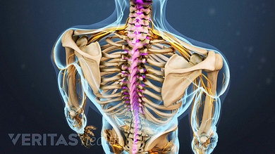
Back Muscles Diagram - Back Muscles Diagram Photos And Premium High Res Pictures Getty Images. The deltoid, teres major, teres minor, infraspinatus, supraspinatus (not shown) and subscapularis muscles (not shown) all extend from the scapula to the humerus and act on the shoulder joint. This muscular system diagram shows the major muscle groups from the back or posterior view. Click on the labels below to find out more about your muscles. To see a muscular system picture from the anterior (front) view click here. Lower back muscle diagram anatomy does degenerative disc disease affect the lower back muscle?
Anatomynote.com found anatomy of back muscles diagram from plenty of anatomical pictures on the internet. It is opposite from the chest, and the vertebral column runs down the back. The human back extends from the buttocks to the posterior portion of the neck and shoulders. Support and protect your spine; Creatine is now proving to be one of the most potent muscle growth accelerators giving excellent muscle mass increase and phenomenal strength increases order yours today.
Back Muscle Diagram For Kids Human Muscle System Muscle Diagram Muscle Anatomy Supraspinatus Muscle The Back Supports The Weight Of The Body Allowing For Flexible Movement While Protecting Vital Organs from i1.wp.com The muscles of your back support your spine, attach your pelvis and shoulders to your trunk, and provide mobility and stability to your trunk and spine. The anatomy of your back muscles can be complex. It is opposite from the chest, and the vertebral column runs down the back. The muscles of the back that work together to support the spine, help keep the body upright and allow twist and bend in many directions. By the way, have you heard about the myth of. The multifidus, a long muscle that travels nearly the entire length of the back.it helps to stabilize and rotate the lower back, and additionally takes some. The part of the nerve that emerges out of the spine is called the nerve root. The muscles, bones, ligaments, and tendons in the back can all be injured and cause back pain.
Deep back muscles superficial back muscles action movements of the shoulder.
This muscle is a major generator of lower back and hip pain, as well as being responsible for complaints of a burning sensation along the posterior superior iliac spine (psis) and sacroiliac joint. To see a muscular system picture from the anterior (front) view click here. The extrinsic (superficial) back muscles, which lie most superficially on the back. How many muscles are in the back? Anatomynote.com found anatomy of back muscles diagram from plenty of anatomical pictures on the internet. Click on the labels below to find out more about your muscles. The back has a total of 40 muscles. Three types of back muscles that help the spine function are extensors, flexors and obliques. Most of the time, back muscle pain is diagnosed then treated with little more than a prescription of rest, painkillers and muscle relaxants. Related posts of back muscles chart muscle anatomy diagram. This muscular system diagram shows the major muscle groups from the back or posterior view. The extensor muscles are attached to back of the spine and enable standing and lifting objects. Anatomy muscles view 12 photos of the anatomy muscles view anatomy muscles view, anatomy of body muscles back view, muscle anatomy anterior view, muscle anatomy back view, muscle anatomy posterior view, human muscles, anatomy muscles view, anatomy of body muscles back view, muscle anatomy anterior view.
The extensor muscles are attached to back of the spine and enable standing and lifting objects. Extrinsic and intrinsic.the back functions are many, such as to house and protect the spinal cord, hold the body and head upright, and adjust the movements of the upper and lower limbs. The back muscles can be three types. We think this is the most useful anatomy picture that you need. The muscles of the back can be arranged into 3 categories based on their location:
Spinal Anatomy And Back Pain from embed.widencdn.net Muscles of lower back diagram. Superficial back muscles, intermediate back muscles and intrinsic back muscles.the intrinsic muscles are named as such because their embryological development begins in the back, oppose to the superficial and intermediate back muscles which develop elsewhere and are therefore classed as extrinsic muscles. The extensor muscles are attached to back of the spine and enable standing and lifting objects. Back to tracking tools main page. Below you'll see diagrams along with the names of the back muscles that may be the cause of your pain. Muscle strain is often the cause of back pain from heavy lifting or vigorous exercise. The pelvis at the bottom of the back and the shoulders at the top of the back give the back. Three types of back muscles that help the spine function are extensors, flexors and obliques.
Lower back muscle diagram anatomy does degenerative disc disease affect the lower back muscle?
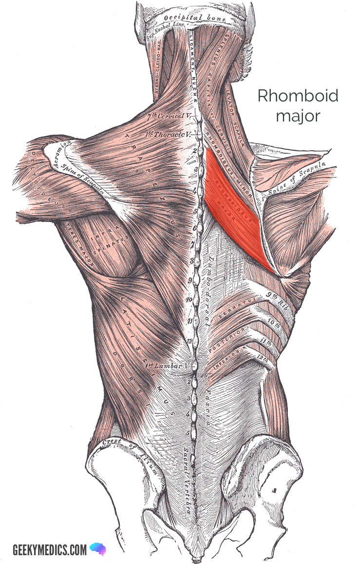
The back muscles enable you to stand up straight; The trapezius and latissimus dorsi muscles connect the upper limb to the vertebral column. Support and protect your spine; Lower back muscle diagram anatomy does degenerative disc disease affect the lower back muscle? Anatomy muscles view 12 photos of the anatomy muscles view anatomy muscles view, anatomy of body muscles back view, muscle anatomy anterior view, muscle anatomy back view, muscle anatomy posterior view, human muscles, anatomy muscles view, anatomy of body muscles back view, muscle anatomy anterior view. The multifidus, a long muscle that travels nearly the entire length of the back.it helps to stabilize and rotate the lower back, and additionally takes some. It comprises the vertebral column (spine) and two compartments of back muscles; Daniel nelson on january 1, 2019 2 comments 🔥! The muscles of the back are a group of strong, paired muscles that lie on the posterior aspect of the trunk they provide movements of the spine, stability to the trunk, as well as the coordination between the movements of the limbs and the back muscles are divided into two large groups: Click on the labels below to find out more about your muscles. This picture also contains humerus, olecranon process of ulna, deep to tendon and so on. As you can see, there are also have a spine of scapula deltoid, triceps brachii, latissimus dorsi. Nerves in your lower back.
The back is the body region between the neck and the gluteal regions. It is opposite from the chest, and the vertebral column runs down the back. For example, some muscles located in the chest also help move the shoulders. By the way, have you heard about the myth of. As you can see, there are also have a spine of scapula deltoid, triceps brachii, latissimus dorsi.
Superficial Back Muscles Anatomy Geeky Medics from geekymedics.com By the way, have you heard about the myth of. The muscles of the back are a group of strong, paired muscles that lie on the posterior aspect of the trunk they provide movements of the spine, stability to the trunk, as well as the coordination between the movements of the limbs and the back muscles are divided into two large groups: There are several different layers of muscles in your back that are often pulling in different and various directions. Deep muscles of the lower back include: These structures work together to support the body, enable a range of movements, and send messages from the. The extensor muscles are attached to back of the spine and enable standing and lifting objects. Creatine research more than a sports supplement read more…. As you can see, there are also have a spine of scapula deltoid, triceps brachii, latissimus dorsi.
Most of the time, back muscle pain is diagnosed then treated with little more than a prescription of rest, painkillers and muscle relaxants.
Back to tracking tools main page. There are several different layers of muscles in your back that are often pulling in different and various directions. Likewise, there are muscles in other parts of the body that help support and move the spine. The multifidus, a long muscle that travels nearly the entire length of the back.it helps to stabilize and rotate the lower back, and additionally takes some. We think this is the most useful anatomy picture that you need. The back consists of the spine, spinal cord, muscles, ligaments, and nerves. Anatomynote.com found anatomy of back muscles diagram from plenty of anatomical pictures on the internet. The extrinsic (superficial) back muscles, which lie most superficially on the back. The back muscles enable you to stand up straight; Muscles of lower back diagram. The muscles of the back that work together to support the spine, help keep the body upright and allow twist and bend in many directions. Related posts of back muscles chart muscle anatomy diagram. The trapezius and latissimus dorsi muscles connect the upper limb to the vertebral column.
Share :
Post a Comment
for "Back Muscles Diagram - Back Muscles Diagram Photos And Premium High Res Pictures Getty Images"


Post a Comment for "Back Muscles Diagram - Back Muscles Diagram Photos And Premium High Res Pictures Getty Images"